Trending
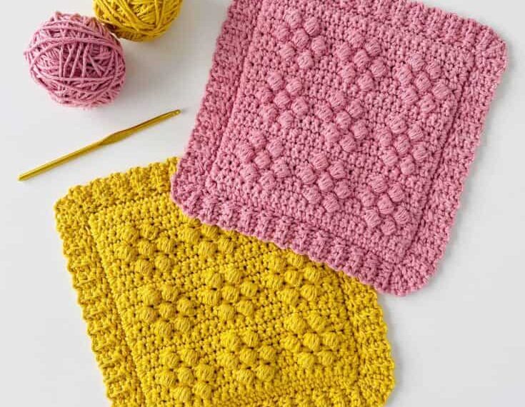
20 Stunning Sugar ‘n Cream Yarn Patterns
In this fast-paced world where you want everything to be on schedule, there is something undeniably therapeutic about…

Discover 30 Cozy Velvet Yarn Crochet Patterns
Remember the time when we were kids, and our body was donned with many crochet wearings like caps,…
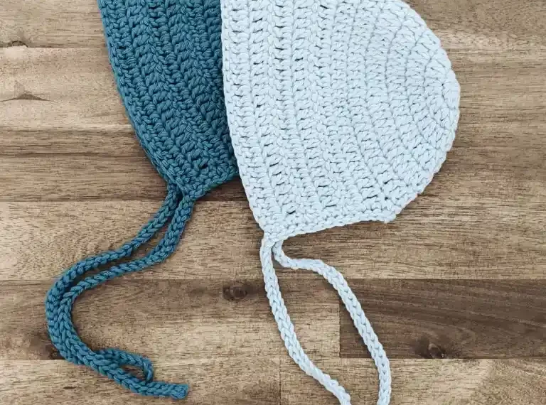
24 Heartwarming Crochet Baby Bonnet Patterns
In the crocheting world, there’s a special place where tradition, artistry, and adorable charm converge—the realm of baby…
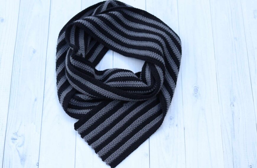
20 Must Try Striped Scarf Crochet Pattern for The Cozy Winter
Winters are meant for hot chocolates and celebrations around the fireplace in the comfiest pajamas and pullovers topped…
Crochet Ruffled Edging/Borders: Full Tutorial
If you are bored of monotonous crochet designs and are looking for a different way to add unique…
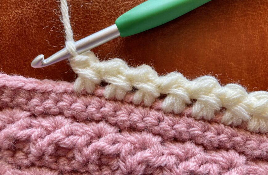
Sedge Stitch Tutorial for Beginners: Step-By-Step Guide
Crocheters now had amazing techniques where they could transform a simple chunky yarn into a beautiful creation, and…
Recent Posts
20 Stunning Sugar ‘n Cream Yarn Patterns
In this fast-paced world where you want everything to be on schedule, there is something undeniably therapeutic about crafting. Using your hands to create something beautiful with your imagination and innovation is a feeling that cannot be explained in words. Enter sugar ‘n cream yarn, a versatile companion for crafters of all stripes. This can bring your creative aspiration into reality, whether you are a crochet connoisseur or new at…
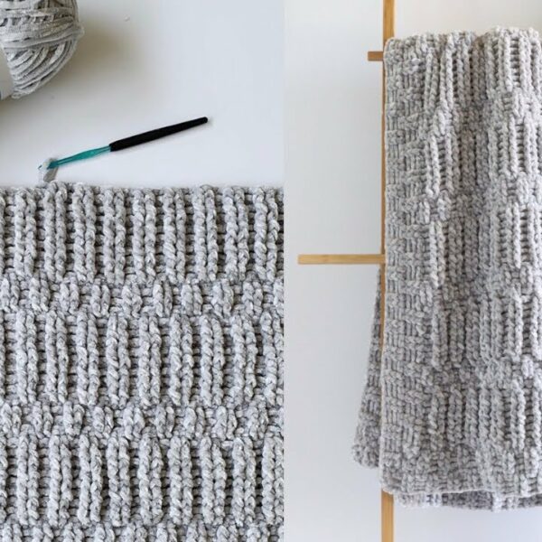
Discover 30 Cozy Velvet Yarn Crochet Patterns
Remember the time when we were kids, and our body was donned with many crochet wearings like caps, socks, sweaters, hand gloves, blankets, and whatnot? Above all, everything felt super…
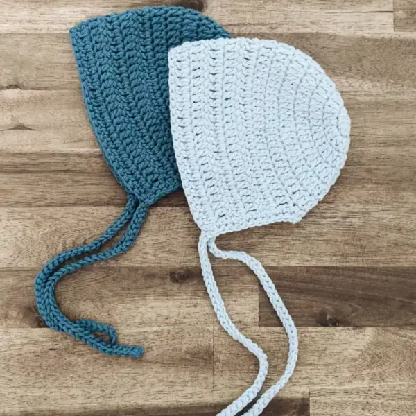
24 Heartwarming Crochet Baby Bonnet Patterns
In the crocheting world, there’s a special place where tradition, artistry, and adorable charm converge—the realm of baby bonnets. These miniature masterpieces hold a timeless allure, preserving the essence of…
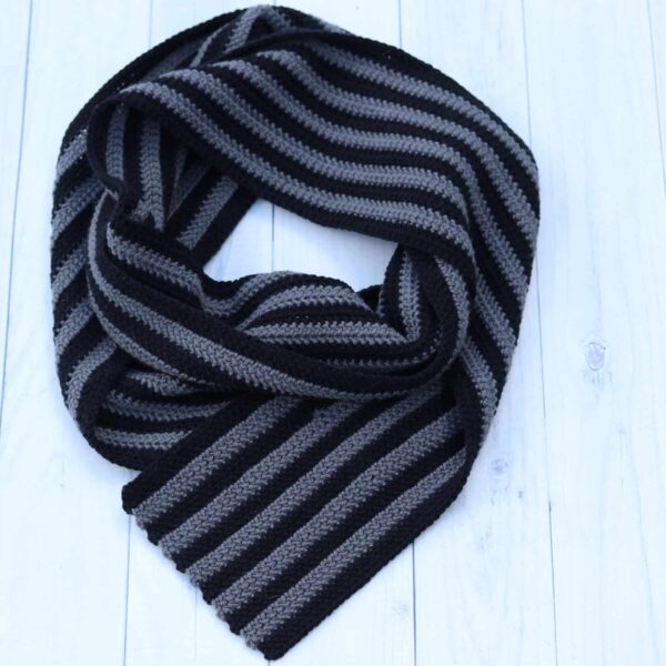
20 Must Try Striped Scarf Crochet Pattern for The Cozy Winter
Winters are meant for hot chocolates and celebrations around the fireplace in the comfiest pajamas and pullovers topped with scarves around the neck. These scarves work as body shields as…